What Is a Screening Mammogram?
A mammogram is an X-ray test of the breast (mammary glands) used to screen for breast abnormalities.
Why Is It Done?
A mammogram is done to help screen for or detect breast cancer. Many small tumors can be seen on a mammogram before they can be felt by a woman or her health care professional.
Mammograms do not prevent breast cancer or reduce a woman's risk of developing cancer. However, regular breast cancer screening can reduce a woman's risk of dying from breast cancer by detecting it in its early stages when it may be easier to treat.
What Is the Difference Between a Screening Mammogram and a Diagnostic Mammogram?
Screening Mammogram
A screening mammogram is a routine, noninvasive X-ray of the breast that looks for potential breast abnormalities. It usually consists of two standard images of each breast.
Screening mammograms are used for early detection of breast cancer when the patient does not have any signs or symptoms of breast disease. According to the American College of Radiology, women at average risk for breast cancer should have a screening mammogram each year beginning at age 40. If you have an increased risk of breast cancer, like family history, your doctor may recommend starting earlier or using a different timeline.
If you are experiencing new breast symptoms, including lumps, pain, skin changes or nipple discharge, it is important to see your health care provider, even if you are not due for your annual mammogram.
Along with offering 3-D screening mammograms at all locations, Memorial Hermann also offers the SmartCurve™ system for 3D screening mammograms at some locations. This technology provides a curved compression surface for a more comfortable patient experience. Your mammography technologist will confirm if you are a candidate for SmartCurve™ during your screening appointment.
Diagnostic Mammogram
A diagnostic mammogram is used to evaluate abnormalities that have already been detected, either through a breast self-exam, a manual exam by your provider or a screening mammogram. Diagnostic mammography is performed to provide radiologists with a more in-depth picture of problems, changes or concerning symptoms. A diagnostic mammogram can also be used for a patient that was previously treated for breast cancer. These X-rays consist of two standard images of each breast, plus more focused images of the area(s) of concern. The additional pictures allow for more accurate and effective characterizations of the symptom or abnormality noted on screening mammogram.
How Do I Prepare for a Screening Mammogram?
If you are still having menstrual periods, you may want to schedule your mammogram within the two weeks following your menstrual period. The procedure may be more comfortable during this time, especially if your breasts become tender before or during your period.
Comparison with prior imaging is key to assessing for change and accurate diagnosis. Please bring a CD and reports of your prior studies if your prior breast health studies were performed outside of Memorial Hermann. If you need us to request your prior films from another facility, please complete a medical release of information form and return it to our facility prior to your appointment. Once received, we will request the films.
On the day of your mammogram, do not use any deodorant, perfume, powders or ointments on your breasts. The residue left on your skin by these substances may interfere with the X-ray images .

Mammogram Reminder

Early detection is the best defense against breast cancer, which is why Memorial Hermann can send you a reminder to schedule your annual screening mammogram.
Remind MeHow Is It Done?
A mammogram is done by a radiology/mammogram technologist. You will need to:
- Remove any jewelry that might interfere with the X-ray picture
- Remove your clothes above the waist; you will be given a cloth or paper gown to wear for the test.
If you are concerned about an area of your breast, show the technologist so that the area can be noted. You usually stand during a breast cancer screening; sometimes you may also be asked to sit or lie down, depending upon the type of X-ray equipment used. One at a time, your breasts will be positioned on a flat plate that will acquire the image. Another plate compresses your breast tissue. Very firm compression is needed to obtain high-quality pictures.
If you are concerned about an area of your breast, show the technologist so that the area can be noted. You usually stand during a breast cancer screening; sometimes you may also be asked to sit or lie down, depending upon the type of X-ray equipment used. One at a time, your breasts will be positioned on a flat plate that will acquire the image. Another plate compresses your breast tissue. Very firm compression is needed to obtain high-quality pictures.
How Long Does It Take?
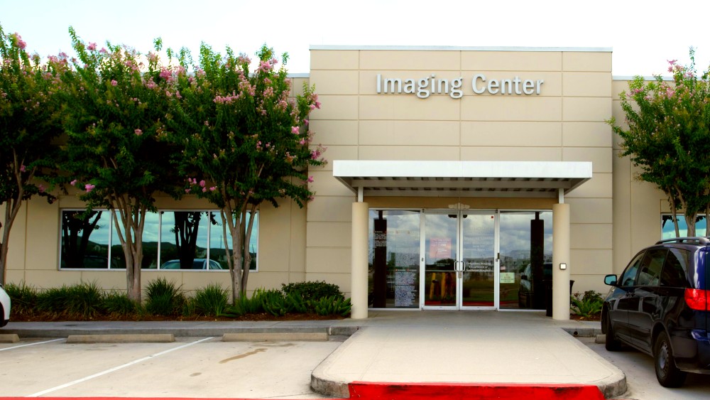
You may be in the imaging center or breast care center for up to an hour; the mammogram itself takes about 10 to 15 minutes.
How Does It Feel?
The X-ray plate will feel cold when you place your breast upon it. Having your breasts flattened and squeezed can be uncomfortable. However, it is necessary to flatten out the breast tissue to obtain the best images.
What Happens After the Test?
The X-ray plate will feel cold when you place your breast upon it. Having your breasts flattened and squeezed can be uncomfortable. However, it is necessary to flatten out the breast tissue to obtain the best images.
Are There Limitations to Mammography?
A mammogram is an important tool to detect breast cancer, but there are limitations based on factors like age and breast density. It is important to speak with your doctor about your personal risk factors, when you should begin screening mammograms and if an annual schedule is best for you.
Make an Appointment
Contact Us
Complete the form below to be connected to our Nurse Navigator – a dedicated registered nurse who specializes in breast health and is available to provide education and resources.


